how do they x ray babies hips
It can cause hip dislocation. All babies are born with spaces between the bones in their skulls.

Degenerative Joint Disease Frog Leg Hip Radiograph Shows Superolateral Joint Space Narrowing Sclerosis Subchondral Cyst A Radiography Osteophyte Radiology
After around 4 to 6 months of age X-rays are the preferred method for evaluating and monitoring hip dysplasia.

. This is called physiologic bow legs. 1RM Bench Press measure of the maximal weight that can be bench pressed with one repetition. Another problem is hip dysplasia where the ball at the end of the femur is loose in the hip socket.
Hip dysplasia is an abnormality of the hip joint where the socket portion does not fully cover the ball portion resulting in an increased risk for joint dislocation. The earlier the condition is treated the better the chance of a successful outcome meaning a hip that appears anatomically normal both during physical examination and on X-ray. Babies who have hip dysplasia are usually born with it but sometimes they develop it later.
Thanks to advances in medicine 90 of babies who have this defect live to. Every year have spina bifida. 10m Beep Test 10m multi-stage walking shuttle test for children with.
They may also send you for some tests or an x-ray. Hip dysplasia may occur at birth or develop in early life. As a child starts walking the bowing might increase a bit and then get better.
The main symptom of colic is localized pain in the abdomen or urinary tract that comes and goes. We would like to show you a description here but the site wont allow us. Treatment for DDH varies between children and depends on its severity.
Babies with DDH can be successfully treated with a special brace. Colic occurs when there is an obstruction. Its considered a normal part of a childs growth and development.
But a child who still has bowlegs at about age three should be evaluated by orthopedic specialist. Mild cases of spina bifida occulta closed neural tube defects not diagnosed during prenatal testing may be detected postnatally using ultrasound or X-ray imaging to look. They may also ask if your child shows any signs of being in pain when they walk and whether any members of your close family have had long-term walking problems.
A diagnostic test that produces images of internal tissues bones and organs that can be used to look at bone structure and alignment. How is hip dysplasia treated in babies. Other diagnostic procedures may include.
Your doctor will probably talk to you and examine you. Children who are treated for hip dysplasia are examined on regular intervals until they are skeletally mature when growth is completed to ensure that normal. Babies with myelomeningocele and closed neural tube defects may have muscle weakness in their feet hips and legs that result in joint deformities first noticed at birth.
They may ask you to keep a diary of your bowel habits where you list what you eat and drink and how often you poo. Regardless it does not typically produce symptoms in babies less than a year old. As the baby grows the space between the bones slowly closes up and disappears and special joints called sutures say.
Over time the bones in the joint rub together causing pain swelling stiffness and reduced movement. Your doctor will recommend the best treatment option for your child. Treatment for hip disorders may include rest medicines physical therapy or surgery including hip replacement.
SOO-churs connect the bones. About 1500 to 2000 babies of the 4 million born in the US. If a physical exam an ultrasound or an X-ray confirm a diagnosis your pediatrician will likely refer you to a pediatric orthopedic specialist for continued care and treatment.
1-RM Tests measure of the maximal weight a subject can lift with one repetition. 1031 production test - concurrent upload 2 TED Talk. This allows the bones to move close up and even overlap as the baby goes through the birth canal.
List of all Tests alphabetical 1 Mile Endurance Run Walk Test complete one mile in the fastest possible time. This can make it harder to walk climb stairs or do other daily activities. Photo and standing-alignment X-ray of a child with bowlegs.
The condition can affect any joint in the body but mostly it affects the knees hips finger joints and big toe. Depending on the cause of the problem they might refer you to a specialist or a continence health professional. Bowlegs refers to a condition in which a persons legs appear bowed bent outward even when the ankles are together.
Your doctor may ask for an ultrasound or X-ray of the hip joint to diagnose DDH. Occasionally one leg may be shorter than the other. It is normal in babies due to their position in the womb.
When babies are born with bow legs its because some of the bones had to rotate twist slightly when they were growing in the womb to fit into the small space.

Pin By Meg Carter On Ortho Hip Dysplasia X Ray Orthopedics
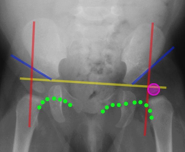
Lines Of The Hip Pediatrics Pediatrics Pediatric Nurse Practitioner Pediatric Radiology

Developmental Dysplasia Of The Hip Ddh Diagnostic Imaging Developmental Dysplasia Of The Hip Diagnostic Imaging Case Study

Pin On X Rays
Pin On درمانی

X Ray Image Of Child Swallowed The Coins For A Medical Diagnosis Medicine Pictures Children Images X Ray Images

Basic Information About Dog Hip Dysplasia Paperblog Dog Hip Dysplasia Hip Dysplasia Canine Hip Dysplasia

Pin On Hip Dysplasia Resources

Hip Dysplasia In Adolescents And Young Adults Hss Hipproblems Hip Dysplasia Hip Problems Hips

Pin On Veterinary Medicine

Pin En Unusual X Rays

Anatomy Pathology Medicine Nursing Radiography Radiologictechnologist Radiology Radiologystudent Instagram Medical Anatomy Radiology Student Radiology

Pin On Nursing 1st Semester

How To Shower After Hip Replacement Surgery Livestrong Com Hip Replacement Surgery Hip Replacement Exercises Hip Brace

This Is An X Ray Of A 2 Week Old Puppy Look At How Far The Bones Have To Grow Before They Become A Proper Bony Joint This Is Why Puppies

Severe Hip Dysplasia In A Boxer The Red Arrows Are Pointing To The Over Growth Of Bone At The Femoral Neck Head The Red Arrow Shades Of Grey Hip Dysplasia

Healthfully Bursitis Hip Hip Workout Hip Replacement

Anatomy And Physiology Anatomy Sacroiliac Joint

Native American Swaddle Hip Dysplasia Baby Developmental Dysplasia Of The Hip Baby Wearing